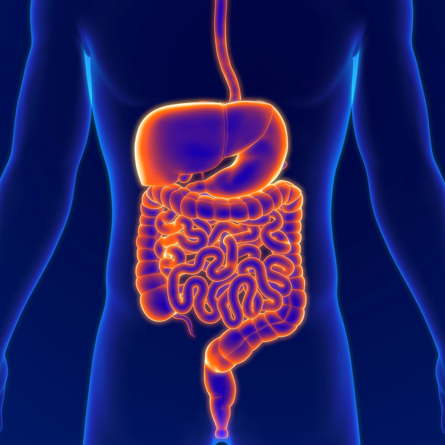
The gut-liver axis refers to the crosstalk between the entire intestine, the resident microbiome and the liver. This crosstalk is fundamental to maintaining homeostasis and human health. A broad and increasing range of literature supports a critical role of a disrupted gut-liver axis in the pathogenesis and progression of liver disease, which makes it a compelling therapeutic target.
Gut-liver axis in health
The gut and the liver interact via their anatomical association. Venous blood from the small and large intestine, spleen and pancreas drains into the portal vein, which provides the majority of the liver’s blood supply. The liver, in tandem, communicates with the intestine via secretion of bile which contains not only digestive enzymes but also important metabolites. The vital bidirectional interaction between these organs is essential for their critical and diverse functions(1). Pathological disruption to this interaction can lead to a variety of disorders and diseases. The gut-liver axis is therefore increasingly recognised as being vital to human health and consists of multiple key components that are further described below and are depicted in Figure 1.
Gut microbiome
The human gastrointestinal tract hosts over 100 trillion microorganisms including bacteria, fungi, viruses and archaea that make up the gut ‘microbiota’. The gut ‘microbiome’ refers to the entire habitat, including the microorganisms, their genes and the surrounding environment, all of which are critical to maintain homeostasis. One of the most important contributions to a diverse gut microbiome in maintaining health is by protection against pathogen colonisation and subsequent infection, recently proven to occur through consumption of the nutrients that bacteria require to colonise a host(2). The gut microbiome is affected by a whole range of lifestyle and environmental factors including diet, physical activity, medications (3) and the circadian rhythm (5).
Intestinal barrier
A functional intestinal barrier is essential for the absorption of nutrients, but also for keeping the trillions of microorganisms contained within the gut lumen and to prevent systemic dissemination where their distant effects may cause harm. It achieves this by having a complex system of cells and ‘tight junction’ between cells that allow different absorption, diffusion and passage of these various elements. Microbial products and metabolites are vital to maintaining the gut barrier and liver health(4). The barrier consists of several intersecting components:
- The most external layer of defence is the mucus, which is where the outer, microbiome-colonised layers interact, while the inner sterile microbe-free layer covers the gut epithelium. More than just a first line of defence, it also serves as a nutrient source and niche for microbiome habitation(5).
- Just below the mucus layer, the gut barrier is formed of a monolayer of epithelial cells. This layer maintains a physical barrier which is dependent on dynamic tight junctions which seal them together; an electrical barrier, as the brush border is negatively charged and opposes a negative charge on the microbiota; and a chemical barrier as a series of antimicrobial peptides are released by epithelial cells(6).
- The next layer of defence is provided by the immune cells of the ‘lamina propria’, which contains several types of innate and adaptive immune cells that all interact and are tightly regulated to ensure that mucosal immunity is maintained.
- The final layer is the gut vascular barrier (GVB) which represents the inner layer of defence that finely regulates the translocation of pathogens, their products and metabolites from the intestinal lumen into and the systemic circulation. Targeting the GVB and the active molecules that drive the vascular connection of the gut-liver-brain axis promises to provide additional therapeutic approaches to diseases affecting organs other than the intestine(7).
The gut microbiome which is found within the intestinal lumen as well as adherent to the gut wall plays a pivotal role in maintaining normal function and homeostasis with all these various components, and especially in the priming and maintenance of gut mucosal immunity (8).
Metabolites
Much of the interaction between the host and the microbiome is mediated by a multitude of soluble factors, which include bacterial products (both beneficial and toxic) and metabolites, that also act as key signalling products. A large array of metabolites drives the crosstalk between the host and its microbiome. Two important ones are short-chain fatty acids (SCFAs) whose production is dependent on the microbiome, and bile acids which are produced by and secreted from the liver into the gut. Others include indoles and other tryptophan metabolites related to the gut-liver-brain axis (9). Trymethylamine N-oxide (TMAO) is produced by intestinal bacteria converting dietary components such as choline or carnitine contained in meat and cheese and has been associated with cardiovascular disease (10).
- Short chain fatty acids (SCFAs) primarily include butyrate, propionate and acetate and are formed in the large intestine by the dietary assimilation of polysaccharides, resistant starch, and fibre. They work as nutrient and energy source for intestinal epithelium and act as precursors for lipogenesis and gluconeogenesis. (11). A decrease in butyrate is observed in several liver diseases and alcohol-related liver injuries (12).
- Bile acids: the liver regulates aspects of gut function by secreting bile, a liquid rich in IgA, bicarbonate, antimicrobial molecules and bile acids. Bile acids are molecules generated in the liver from cholesterol, transported as primary conjugated bile acids into the biliary system, which drains them into the small intestine. There, they can be further metabolised by the microbiome into secondary bile acids. Beyond their role as osmotic drivers of bile flow and detergents for lipid digestion and absorption, they regulate hepatic metabolism, shape the gut microbiome composition and function, and sustain intestinal barrier integrity. The crosstalk between bile acids and gut microbiome is a two-way interaction with both influencing each other’s function (13). Bile acids exert bacteriostatic effects directly via their detergent properties. Indirectly this occurs via activation of the bile acid farnesoid X receptor (FXR) to stimulate the production of antimicrobial molecules in intestinal epithelial cells and to maintain the gut vascular barrier (14). Bile acids have evolved not only as crucial factors affecting the intestinal microbiome, but may also contribute to longevity as suggested by a study in centenarians (15).
In summary, in a healthy state, the gut microbiome resides in the gut lumen, yet its metabolites act locally and systemically to maintain homeostasis of the gut–liver axis. Disruption of these various components and their interactions can contribute to liver disease and disorders beyond the liver such as neurological, cardiovascular disease and unhealthy aging.
Gut-liver axis in chronic liver disease
The crosstalk between the gut microbiome, its derived metabolites, the immune system, and the liver plays a key role in the pathogenesis of chronic liver diseases (4). Patients with cirrhosis are predisposed to small intestinal bacterial overgrowth (16) due to liver tissue scarring and decreased bile flow. Gut barrier dysfunction leads to the influx of ‘pathogen-associated molecular patterns’ (PAMPs) and microbiome-derived metabolites to the liver, that also trigger and propagate hepatic inflammation(6).
Gut dysbiosis is very broadly described as alterations in microbial composition, changes in microbial metabolic activity, and/or changes in their distribution within the entire gut, which includes the oral cavity (17). More recently, a more refined description has been proposed to include a distinct or altered microbial ecological state that is causally linked to the manifestation, diagnosis or treatment of a disease (18). This emphasises the need to better understand cause-and-effect and underpins the requirement for more robust research including relevant disease models and well-designed clinical trials (1). Dysbiosis and gut barrier dysfunction are directly involved in the pathogenesis of stable cirrhosis, where there is liver scarring but liver function is maintained.
Figure 1. Relationship of the gut microbiome to other components of the intestinal barrier and the gut-liver axis in chronic liver disease (figure originating from (8))
The types and severity of complications in decompensated cirrhosis, such as bacterial infections, encephalopathy, extrahepatic organ failure and progression to acute on chronic liver failure(19) (see Figure 1), are increasingly linked to gut and even oral dysbiosis(20, 21). Differences in bacterial, viral and fungal compartments are seen in cirrhosis at all stages of severity and are most evident for alcohol-related and steatotic liver disease (22). Excessive alcohol intake has a direct and deleterious impact on the gut microbiome, independent to liver inflammation and cirrhosis that alcohol causes (23). Dysbiosis alters the metabolism of intestinal bile acids, with less conversion of primary to secondary bile acids, which is toxic to the gut wall. Changes in luminal bile acids and in FXR activity are connected to gut barrier dysfunction and bacterial translocation. Total and conjugated serum bile acids correlate with liver disease severity (7).
Manipulation of intestinal microbial communities has been shown to modulate and potentially treat liver disease: these therapeutic effects can be transmitted with faecal microbiota in experimental models,(24, 25) and an improvement in metabolic and liver pathologies has been reported after faecal microbiota transplantation from healthy donors (26). Non faecal transplant-based approaches such as dietary manipulation, pre-/pro-/synbiotics and other gut targeted therapies need further evaluation to fully harness their potential in the context of not only liver diseases (6, 27) but other chronic conditions. Taking all this into account, targeting the gut-liver axis for the treatment of chronic liver disease represents a novel and exciting therapeutic avenue.
Conflicts of Interest: the authors have none to declare relating to this work.
References
1. Tripathi A, Debelius J, Brenner DA, Karin M, Loomba R, Schnabl B, et al. The gut-liver axis and the intersection with the microbiome. Nat Rev Gastroenterol Hepatol. 2018;15(7):397-411.
2. Spragge F, Bakkeren E, Jahn MT, E BNA, Pearson CF, Wang X, et al. Microbiome diversity protects against pathogens by nutrient blocking. Science. 2023;382(6676):eadj3502.
3. Jackson MA, Verdi S, Maxan ME, Shin CM, Zierer J, Bowyer RCE, et al. Gut microbiota associations with common diseases and prescription medications in a population-based cohort. Nat Commun. 2018;9(1):2655.
4. Tilg H, Adolph TE, Trauner M. Gut-liver axis: Pathophysiological concepts and clinical implications. Cell Metab. 2022;34(11):1700-18.
5. Derrien M, Van Baarlen P, Hooiveld G, Norin E, Muller M, de Vos WM. Modulation of Mucosal Immune Response, Tolerance, and Proliferation in Mice Colonized by the Mucin-Degrader Akkermansia muciniphila. Front Microbiol. 2011;2:166.
6. Albillos A, de Gottardi A, Rescigno M. The gut-liver axis in liver disease: Pathophysiological basis for therapy. J Hepatol. 2020;72(3):558-77.
7. Sorribas M, Jakob MO, Yilmaz B, Li H, Stutz D, Noser Y, et al. FXR modulates the gut-vascular barrier by regulating the entry sites for bacterial translocation in experimental cirrhosis. J Hepatol. 2019;71(6):1126-40.
8. Woodhouse C, Singanayagam A, Patel VC. Modulating the gut-liver axis and the pivotal role of the faecal microbiome in cirrhosis. Clin Med (Lond). 2020;20(5):493-500.
9. Hendrikx T, Schnabl B. Indoles: metabolites produced by intestinal bacteria capable of controlling liver disease manifestation. J Intern Med. 2019;286(1):32-40.
10. Schiattarella GG, Sannino A, Toscano E, Giugliano G, Gargiulo G, Franzone A, et al. Gut microbe-generated metabolite trimethylamine-N-oxide as cardiovascular risk biomarker: a systematic review and dose-response meta-analysis. Eur Heart J. 2017;38(39):2948-56.
11. Martin-Gallausiaux C, Marinelli L, Blottiere HM, Larraufie P, Lapaque N. SCFA: mechanisms and functional importance in the gut. Proc Nutr Soc. 2021;80(1):37-49.
12. Juanola O, Ferrusquía-Acosta J, García-Villalba R, Zapater P, Magaz M, Marín A, et al. Circulating levels of butyrate are inversely related to portal hypertension, endotoxemia, and systemic inflammation in patients with cirrhosis. FASEB J. 2019;33(10):11595-605.
13. Wahlström A, Sayin SI, Marschall HU, Bäckhed F. Intestinal Crosstalk between Bile Acids and Microbiota and Its Impact on Host Metabolism. Cell Metab. 2016;24(1):41-50.
14. Hsu CL, Schnabl B. The gut-liver axis and gut microbiota in health and liver disease. Nat Rev Microbiol. 2023;21(11):719-33.
15. Sato Y, Atarashi K, Plichta DR, Arai Y, Sasajima S, Kearney SM, et al. Novel bile acid biosynthetic pathways are enriched in the microbiome of centenarians. Nature. 2021;599(7885):458-64.
16. Bauer TM, Schwacha H, Steinbruckner B, Brinkmann FE, Ditzen AK, Aponte JJ, et al. Small intestinal bacterial overgrowth in human cirrhosis is associated with systemic endotoxemia. Am J Gastroenterol. 2002;97(9):2364-70.
17. Petersen C, Round JL. Defining dysbiosis and its influence on host immunity and disease. Cell Microbiol. 2014;16(7):1024-33.
18. Levy M, Kolodziejczyk AA, Thaiss CA, Elinav E. Dysbiosis and the immune system. Nat Rev Immunol. 2017;17(4):219-32.
19. Philips CA, Augustine P. Gut Barrier and Microbiota in Cirrhosis. J Clin Exp Hepatol. 2022;12(2):625-38.
20. Albuquerque-Souza E, Sahingur SE. Periodontitis, chronic liver diseases, and the emerging oral-gut-liver axis. Periodontol 2000. 2022;89(1):125-41.
21. Acharya C, Sahingur SE, Bajaj JS. Microbiota, cirrhosis, and the emerging oral-gut-liver axis. JCI Insight. 2017;2(19).
22. Bajaj JS, Khoruts A. Microbiota changes and intestinal microbiota transplantation in liver diseases and cirrhosis. J Hepatol. 2020;72(5):1003-27.
23. Engen PA, Green SJ, Voigt RM, Forsyth CB, Keshavarzian A. The Gastrointestinal Microbiome: Alcohol Effects on the Composition of Intestinal Microbiota. Alcohol Res. 2015;37(2):223-36.
24. Henao-Mejia J, Elinav E, Jin C, Hao L, Mehal WZ, Strowig T, et al. Inflammasome-mediated dysbiosis regulates progression of NAFLD and obesity. Nature. 2012;482(7384):179-85.
25. Llopis M, Cassard AM, Wrzosek L, Boschat L, Bruneau A, Ferrere G, et al. Intestinal microbiota contributes to individual susceptibility to alcoholic liver disease. Gut. 2016;65(5):830-9.
26. Philips CA, Ahamed R, Rajesh S, Abduljaleel JKP, Augustine P. Long-term Outcomes of Stool Transplant in Alcohol-associated Hepatitis-Analysis of Clinical Outcomes, Relapse, Gut Microbiota and Comparisons with Standard Care. J Clin Exp Hepatol. 2022;12(4):1124-32.
27. Kaufmann B, Seyfried N, Hartmann D, Hartmann P. Probiotics, prebiotics, and synbiotics in nonalcoholic fatty liver disease and alcohol-associated liver disease. Am J Physiol Gastrointest Liver Physiol. 2023;325(1):G42-G61.


